

 USA
 Australia
 Canada
 India
 Malaysia
 UK
 New Zealand
 España
 France
 Indonesia
 Italia
 México
 Deutschland
 中国
 香港
 日本
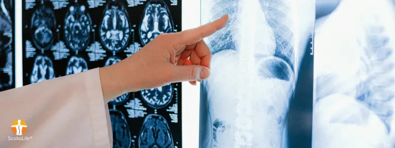
Scoliosis X-rays are safer than the old cancer studies suggest, but the number of films over four to six years of growth is something families can influence. Here is what the evidence shows and how to keep the count low without losing track of the curve.
If your child has scoliosis, X-rays become part of life for several years: a baseline film at diagnosis, follow-up films through the growth spurt, in-brace and out-of-brace films if a brace is prescribed. Somewhere along the way most parents ask the quiet question they are slightly embarrassed to raise: is all this radiation safe?
The honest answer sits between blanket reassurance and alarm. The studies that found higher cancer rates in scoliosis patients are real, but they measured a very different kind of X-ray from the one your child receives today, and the number of films a child accumulates is something families can genuinely influence. Here is what the evidence shows, how many X-rays monitoring really involves, which films are worth having, and how to keep the count low without losing track of the curve.
Where the worry comes from: the historic cohorts
The concern is not a myth. The largest body of evidence is the United States Scoliosis Cohort: roughly 5,500 women diagnosed between 1912 and 1965 who received an average of about 23 spinal radiographs each. In the 2010 follow-up by Ronckers and colleagues, breast cancer mortality was about 1.7 times that of the general population, and the risk rose with the number of films. In Denmark, Simony and colleagues followed 211 adolescents treated between 1983 and 1990, with about 16 radiographs each, and reported a cancer rate several times higher than expected. That finding rests on only nine cases, and the cohort’s estimated cumulative dose was low (about 1.6 mSv on average), which has led researchers to ask whether factors other than radiation also contribute.
A 2025 systematic review in Spine Deformity examined seven such cohorts, and its conclusion is the one to hold on to: the excess risk is mainly breast cancer in women exposed before 1976, with front-to-back (anteroposterior) films at doses far above current practice. The authors judge that modern techniques probably reduce the risk substantially, but note that no cohort imaged with today’s equipment has yet been followed long enough to prove it. That is honest uncertainty, and it argues for using X-rays wisely rather than avoiding them.
How much radiation is in a scoliosis X-ray today
Three changes explain most of the gap between those cohorts and your child’s next film. First, projection: the review estimates that the breast dose from a front-to-back full-spine film fell from about 0.78 cGy in the 1940s and 1950s to about 0.125 cGy by the late 1980s, while a back-to-front (posteroanterior, or PA) film delivers roughly 0.003 cGy, more than forty times lower, because the spine and ribs shield the breast tissue. Second, digital detectors need far less exposure than film. Third, slit-scanning systems such as EOS, which the SOSORT consensus describes as reducing spine doses to about one sixth to one ninth of a standard examination.
In everyday units, a 2020 Danish study put a conventional full-spine examination (front and side views) at about 0.55 mSv and a standard EOS examination at about 0.22 mSv; a Hong Kong study of adolescents measured the PA view alone at about 0.07 mSv with digital radiography and about 0.003 mSv in EOS micro-dose mode. Natural background radiation is roughly 2 to 3 mSv a year, so a single modern full-spine examination sits somewhere between a few days and a few months of background exposure, depending on the equipment and views. The risk from any one film is very small; what is worth managing is the total count over four to six years of growth, and the type of film taken.
How many X-rays does scoliosis monitoring actually involve?
Estimates of 10 to 50 examinations across adolescence have been quoted, and the spread mostly reflects how much protocols differ between clinics. The 2012 consensus of SOSORT, the international society for conservative scoliosis treatment, set out a deliberately restrained schedule for idiopathic scoliosis under observation or non-surgical treatment:
- Juvenile scoliosis (ages 6 to 12): a film about every 6 months, because curves can move quickly before puberty.
- Adolescent scoliosis at Risser 0 to 3 (growth under way): about every 12 months; at Risser 4 to 5 (growth nearly complete): about every 18 months; after growth: about every 24 months during surveillance.
- Clinical visits every 3 to 6 months during growth, with a film brought forward only if the scoliometer reading or visible trunk asymmetry changes.
- A side (lateral) view at the first assessment, not at every follow-up, unless the sagittal profile appears to be changing.
- PA rather than AP projection. CT is reserved for surgical planning and MRI for suspected neurological causes.
Risser is a 0 to 5 grade of how much of the pelvic growth plate has turned to bone, read from the same film as the curve, and one of the main ways clinicians judge how much growth is left. If your child wears a brace, expect a few additional films: one in the brace soon after fitting, to confirm that it is actually correcting the curve, and periodic out-of-brace films at intervals that vary from 3 to 6 months between clinics.
As an illustration, a girl diagnosed at 11 and followed to Risser 4 at about 15 would accumulate roughly 6 to 8 films on a SOSORT-style schedule and 12 to 18 on a routine 3- to 4-monthly schedule, before any bracing films. The protocol, not the diagnosis, decides most of that difference.
Four ways X-rays get wasted
A surprising share of a child’s imaging never changes a decision. The common culprits:
- Repeat films when changing clinics. A full-spine film taken in the last 3 to 4 months rarely needs repeating. Ask for the images in DICOM format, not just the written report, and bring them to any second opinion.
- A side view at every visit. It roughly doubles the dose of a visit and, for most idiopathic curves, adds little once the baseline sagittal profile is known.
- AP instead of PA. Facing the X-ray tube rather than the detector puts breast and thyroid tissue in the primary beam. Many departments default to PA for spines, but ask.
- Films with no question attached. A scan ordered “to see how things are going” when no treatment change would follow, whatever the result, is a film your child did not need. The same Danish study found that a single additional CT can double a patient’s total imaging dose.
Low-dose options worth asking about
EOS is a slit-scanning system that captures standing front and side views of the whole spine simultaneously at a dose well below conventional radiography, with a micro-dose setting for follow-up films where fine detail matters less. In Singapore, KK Women’s and Children’s Hospital offers EOS imaging, and private centres increasingly run low-dose digital protocols. Availability varies, so ask which system will be used, whether a paediatric protocol is in place, and whether the side view is needed on this visit. A child who knows to stand still for the few seconds of the scan also avoids a repeat exposure for a blurred image.
Monitoring between X-rays without radiation
A restrained X-ray schedule is safe because a lot can be measured without radiation. A 2025 meta-analysis in BMC Musculoskeletal Disorders, covering 56 studies and 4,774 patients, found that spinal ultrasound correlates strongly with the radiographic Cobb angle (r of about 0.9) and that surface topography, which maps the shape of the back optically, also correlates well (r above 0.8). Neither yet replaces the X-ray for diagnosis or major treatment decisions, but both are credible tools for tracking a curve between films.
The simplest tool is the scoliometer, the small inclinometer placed across the back during a forward bend. SOSORT’s consensus makes it the gatekeeper: a stable reading supports waiting for the scheduled film, while a clear change justifies bringing it forward. Monthly readings, standardised posture photographs and a record of height velocity, since curves move fastest during the peak growth spurt, give a clinician most of what is needed to judge whether an X-ray now would change anything. Our articles on monitoring mistakes between visits and the scoliosis growth window cover the practical routine, and the free Scoliometer app lets families log readings at home.
Dr Kevin Lau’s perspective: every X-ray should answer a question
What follows is a clinical perspective from ScolioLife®, not a trial finding. Over 25 years of non-surgical scoliosis care, Dr Kevin Lau has come to treat imaging as a budget to be spent deliberately. Every film in a child’s plan should answer a specific question whose answer would change what is done next: has the curve crossed a threshold that changes treatment intensity, is the brace correcting the curve, has growth finished so the plan can be relaxed. If no decision hangs on the result, the film waits.
Two films are rarely worth skipping. The first is a good baseline: a standing PA full-spine film read for Cobb angle, vertebral rotation, Risser grade and sagittal balance all at once, so one exposure yields the whole picture. The second, for braced children, is the first in-brace film, the only direct evidence that a brace is doing its job; a brace that is not correcting the curve needs adjusting now, not at the next annual review. After those two, the question at every visit is whether scoliometer readings, photographs and growth measurements are stable. In the ScolioLife® System these are recorded at each visit and between visits through the ScolioTrack app, and an X-ray is brought forward when they move rather than because a calendar interval has elapsed. Families receive a written imaging plan tied to their child’s growth stage, so nobody is surprised by a film and nobody repeats one that already exists.
Questions to ask before your child’s next X-ray
- What decision will this film inform, and what would change if it were not taken today?
- Is a side view needed this time, or only the front view?
- Will it be taken PA (back to the tube) rather than AP?
- Is a low-dose or EOS system available, and is a paediatric protocol in use?
- What scoliometer reading or visible change should prompt us to come back before the next scheduled film?
Frequently asked questions
Is a scoliosis X-ray safe for a child?
The radiation from a single modern full-spine X-ray is low, roughly equivalent to days to a few months of natural background exposure depending on the equipment and views. The studies that linked scoliosis imaging to cancer involved much older techniques and far higher doses. The sensible aim is not to avoid X-rays but to take only those that will change a decision.
How often should a child with scoliosis have an X-ray?
The SOSORT consensus suggests about every 12 months while growth is under way (Risser 0 to 3), about every 18 months as growth finishes (Risser 4 to 5) and about every 6 months for juvenile scoliosis, with an earlier film only if the scoliometer reading or visible asymmetry changes. Braced children usually need a few extra films, including one in the brace after fitting.
Can scoliosis be monitored without X-rays?
Between X-rays, yes. Scoliometer readings, standardised photographs, height tracking, surface topography and spinal ultrasound all track a curve without radiation, and recent research shows ultrasound and topography correlate strongly with the Cobb angle. None yet replaces the X-ray for diagnosis or major treatment decisions.
What is EOS imaging and should I ask for it?
EOS is a low-dose scanning system that takes standing front and side images of the whole spine at the same time, at a fraction of the dose of conventional radiography, with an even lower micro-dose mode for follow-up. It is available at a growing number of hospitals, including KK Women’s and Children’s Hospital in Singapore, and is a reasonable thing to ask for, especially for repeat films.
Should I bring old X-rays to a new clinic?
Yes, and bring the actual images rather than only the report. A film taken within the last few months can usually be reused, avoiding an unnecessary exposure, and a series of older films shows how the curve has behaved over time, which is often more informative than any single image.
Take the next step
If you would like a second look at your child’s existing X-rays before deciding on the next step, ScolioLife® reviews films from any clinic and builds a written monitoring and treatment plan around your child’s growth stage. Families are seen at the Singapore clinic on Orchard Road, as well as in Kuala Lumpur and Surabaya, with online reviews of existing images for families further afield. Book a consultation or learn more about the ScolioLife® scoliosis programme.
More articles

Does Scoliosis Make You Shorter? How Much Height a Curve Costs, and What Can Be Regained
November 3, 2026
Is Swimming Good for Scoliosis? What the Research Shows, and What the Pool Can and Cannot Do
October 26, 2026
Night-Time vs Full-Time Scoliosis Brace: How to Choose the Right Protocol for Your Child
October 23, 2026